
Slika:Human brain anterior-inferior view description.JPG
Human_brain_anterior-inferior_view_description.JPG (330 × 475 točk, velikost datoteke: 31 KB, MIME-vrsta: image/jpeg)
Spodaj prikazane informacije so s tamkajšnje opisne strani.
Povzetek
| Opis |
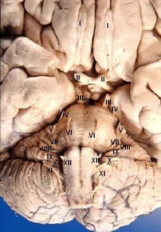
Human brain - anterior-inferior view - cerebral nerves I. N. olfactorius - The fila olfactoria (approximately 20 nerve rootlets on each side) comprise the Olfactory nerves (I) which run from the olfactory receptors in the nasal cavity to the olfactory bulb. II. N. opticus - The Optic nerves (II) are large & join at the midline to form the optic chiasm, then continue laterally as the optic tracts. The optic nerve is not a true nerve but rather a CNS tract. III. N. oculomotorius - The Oculomotor nerves (III) emerge from a depression in the midbrain, the interpeduncular fossa, just caudal to the optic chiasm. IV. N. trochlearis - The Trochlear nerves (IV) are small & are seen on the lateral surface of the midbrain. They are the only nerves which exit from the posterior side of the brain. V. N. trigeminus - The Trigeminal nerves (V) are large and emanate from the lateral surface of the pons. VI. N. abducens - The Abducens nerves (VI) exit near the midline from the inferior pontine sulcus which separates the pons from the medulla. VII. N. facialis - Moving laterally in the inferior pontine sulcus, the Facial nerves (VII) can be seen. VIII. N. vestibulocochlearis - Slightly lateral to the facial nerve is the Vestibulocochlear nerve (VIII). IX. N. glossopharyngeus - The postolivary sulcus is a groove running rostrocaudally on the lateral surface of the medulla. From this sulcus pass the small Glossopharyngeal nerves (IX) rostrally and... X. N. vagus - ...the much larger Vagus nerves (X) caudally. XI. N. accessorius - The Spinal Accessory nerves (XI) exit the cervical cord then pass rostrally through the foramen magnum to exit the cranial vault with the lossopharyngeal and vagus nerves. XII. N. hypoglossus - The Hypoglossal nerves (XII) exit the medulla via the preolivary sulcus.
|
| Datum | |
| Vir | http://www.healcentral.org/healapp/showMetadata?metadataId=40566 (Internet Archive of file description page) |
| Avtor |
John A Beal, PhD Dep't. of Cellular Biology & Anatomy, Louisiana State University Health Sciences Center Shreveport |
| Dovoljenje (Nadaljnja uporaba datoteke) |
CC-BY |
| Druge različice |
 |
Licenca
- Dovoljeno vam je:
- deljenje – reproducirati, distribuirati in javno priobčevati delo
- predelava – predelati delo
- Pod naslednjimi pogoji:
- priznanje avtorstva – Navesti morate ustrezno avtorstvo, povezavo do licence in morebitne spremembe. To lahko storite na kakršen koli primeren način, vendar ne na način, ki bi nakazoval, da dajalec licence podpira vas ali vašo uporabo dela.
To sliko, prvotno objavljeno na http://www.healcentral.org/healapp/showMetadata?metadataId=40566, je 25. septembra 2013 pregledal administrator ali pregledovalec Eleassar in potrdil, da je bila takrat tam na razpolago pod navedeno licenco.
|
Zgodovina datoteke
Kliknite datum in čas za ogled datoteke, ki je bila takrat naložena.
| Datum in čas | Sličica | Velikost | Uporabnik | Komentar | |
|---|---|---|---|---|---|
| trenutno | 20:22, 24. junij 2006 | | 330 × 475 (31 KB) | Patho | {{Information| |Description='''Human brain - anterior-inferior view - cerebral nerves''' I. N. olfactorius - The fila olfactoria (approximately 20 nerve rootlets on each side) comprise the Olfactory nerves (I) which run from the olfactory receptors in |
Uporaba datoteke
Datoteka je del naslednje 1 strani slovenske Wikipedije (strani drugih projektov niso navedene):
Globalna uporaba datoteke
To datoteko uporabljajo tudi naslednji vikiji:
- Uporaba na ar.wikipedia.org
- Uporaba na azb.wikipedia.org
- Uporaba na az.wikipedia.org
- Uporaba na bs.wikipedia.org
- Uporaba na de.wikipedia.org
- Uporaba na de.wikibooks.org
- Uporaba na en.wikipedia.org
- Cranial nerves
- User talk:Hovea
- User talk:Wouterstomp
- User talk:Nephron
- User talk:NCurse
- User talk:Robotsintrouble
- User talk:Was a bee
- User talk:Bloomingdedalus
- User talk:Read-write-services
- User talk:Attys
- User talk:Bakerstmd
- User talk:Manfi
- User talk:Mikepascoe
- User talk:Vokesk
- User talk:Neuraxıs
- User talk:Cmungall
- User talk:Qxukhgiels
- User talk:DocElisa
- User talk:JakobSteenberg/Archives/1
- User talk:Meteor sandwich yum
- User talk:Slashedone
- User talk:Anindya07
- User talk:Mattimussi
- User talk:Jelly Bean MD
- User talk:Benrusholme
- User talk:Brad.w.english
- User talk:Krsna ss
- User talk:Tom (LT)/Archive 4
- User talk:Dregon131
- User talk:Sapientia42
- User talk:Tilifa Ocaufa
- User talk:Snow Rise/Archive 6
- User talk:Arcadian
- User talk:Outofbattery
- User talk:Anatomyczar
- Wikipedia:WikiProject Anatomy/Newsletter/4
- User talk:Supravibhatsupravi
- User talk:Athikhun.suw
- User talk:CFCF/Archive 6
- User talk:Lewisskinner/ArchiveJul 2015
- User talk:Tyrol5/Archive 6
- User talk:CFCF/Archive 7
- User talk:DiverDave/Archive 5
Oglejte si globalno uporabo te datoteke.