
Slika:Brain - Lobes.png
Višja ločljivost ni na voljo.
Brain_-_Lobes.png (701 × 487 točk, velikost datoteke: 360 KB, MIME-vrsta: image/png)
Spodaj prikazane informacije so s tamkajšnje opisne strani.
| Opis |
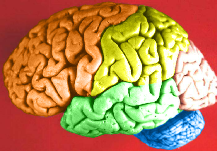
Human brain lateral view - Lobes
|
| Datum | (UTC) |
| Vir | Human_brain_lateral_view_description.JPG |
| Avtor | Dep't. of Cellular Biology & Anatomy, Louisiana State University Health Sciences Center Shreveport |
| Dovoljenje (Nadaljnja uporaba datoteke) |
CC-BY |
| Druge različice |
| Ta slika je digitalno obdelana. Opravljene so bile naslednje spremembe: Hemispheres in color.. Izvirnik je na voljo tukaj: Human brain lateral view description.JPG. Spremembe je opravil uporabnik DavoO.
|
Licenca
Jaz, imetnik avtorskih pravic na tem delu, ga objavljam pod naslednjo licenco:
Datoteka je objavljena pod licenco Creative Commons Priznanje avtorstva 2.5 Generična.
- Dovoljeno vam je:
- deljenje – reproducirati, distribuirati in javno priobčevati delo
- predelava – predelati delo
- Pod naslednjimi pogoji:
- priznanje avtorstva – Navesti morate ustrezno avtorstvo, povezavo do licence in morebitne spremembe. To lahko storite na kakršen koli primeren način, vendar ne na način, ki bi nakazoval, da dajalec licence podpira vas ali vašo uporabo dela.
The following refers to the original source file, not this derivative version.
To sliko, prvotno objavljeno na https://web.archive.org/web/20110514023714/http://www.healcentral.org/healapp/showMetadata?metadataId=40566, je 1. novembra 2013 pregledal administrator ali pregledovalec Avenue in potrdil, da je bila takrat tam na razpolago pod navedeno licenco.
|
Izvorni dnevniški zapis naložitve
This image is a derivative work of the following images:
- File:Human_brain_lateral_view_description.JPG licensed with Cc-by-2.5
- 2006-06-20T13:58:22Z Patho 701x487 (50176 Bytes) {{Information| |Description='''Human brain lateral view - Lobes''' # Lobus frontalis # Lobus parietalis # Lobus temporalis # Lobus occipitalis # Sulcus lateralis # Sulcus centralis # Sulcus parietooccipitalis # Incisura preo
- 2006-06-20T13:54:13Z Patho 701x487 (49891 Bytes) Auf eine alte Version zurückgesetzt
- 2006-06-20T13:51:38Z Patho 701x487 (50074 Bytes) {{Information| |Description='''Human brain lateral view - Lobes''' # Lobus frontalis # Lobus parietalis # Lobus temporalis # Lobus occipitalis # Sulcus lateralis # Sulcus centralis # Sulcus parietooccipitalis # Incisura preo
- 2006-06-20T13:28:44Z Patho 701x487 (49891 Bytes) {{Information| |Description='''Human brain lateral view''' # Lobus frontalis # Lobus parietalis # Lobus temporalis # Lobus occipitalis # sulcus lateralis # Sulcis centralis # Sulcus parietooccipitalis # Incisura preoccipital
Uploaded with derivativeFX
Zgodovina datoteke
Kliknite datum in čas za ogled datoteke, ki je bila takrat naložena.
| Datum in čas | Sličica | Velikost | Uporabnik | Komentar | |
|---|---|---|---|---|---|
| trenutno | 00:11, 24. februar 2009 | | 701 × 487 (360 KB) | DavoO | {{Information |Description='''Human brain lateral view - Lobes''' # Lobus frontalis # Lobus parietalis # Lobus temporalis # Lobus occipitalis # Sulcus lateralis # Sulcus centralis # Sulcus parietooccipitalis # Incisura preoccipitalis # Polus frontalis # |
Uporaba datoteke
Datoteka je del naslednje 1 strani slovenske Wikipedije (strani drugih projektov niso navedene):
Globalna uporaba datoteke
To datoteko uporabljajo tudi naslednji vikiji:
- Uporaba na cs.wikipedia.org
- Uporaba na en.wikipedia.org
- Talk:Alcohol intoxication
- Talk:LSD
- Talk:Scopolamine
- Talk:Qigong
- Talk:Recreational drug use
- Talk:Psilocybin
- Talk:Phenomenology (philosophy)
- Talk:Alcohol (chemistry)
- Talk:Timothy Leary
- Talk:Psilocybe cubensis
- Talk:Nitrous oxide
- Talk:Atropine
- Talk:Out-of-body experience
- Talk:The Doors of Perception
- Talk:Carlos Castaneda
- Talk:Ganzfeld experiment
- Talk:Meditation
- Talk:Zen
- Talk:Hysteria
- Talk:Peyote
- Talk:Hashish
- Talk:Ketamine
- Talk:Coca
- Talk:Hippie
- Talk:Spirituality
- Talk:Mantra
- Talk:N,N-Dimethyltryptamine
- Talk:Amanita muscaria
- Talk:Hookah
- Talk:Autogenic training
- Talk:Psychonautics
- Talk:Hypnosis
- Talk:Dipropyltryptamine
- Talk:Ergot
- Talk:Phencyclidine
- Talk:Anadenanthera peregrina
- Talk:Shamanism
- Talk:Mescaline
- Talk:Diphenhydramine
- Talk:Salvinorin A
- Talk:Human Potential Movement
- Talk:Argyreia nervosa
- Talk:Terence McKenna
- Talk:Psilocybin mushroom
- Talk:Atropa belladonna
- Talk:Hypnagogia
- Talk:Kundalini yoga
- Talk:DiPT
- Talk:Dreamachine
Oglejte si globalno uporabo te datoteke.